
An Illustration of Brain Surgery
by Garni Barkhoudarian
We are pleased to announce the publication of our latest article in the December 2017 issue of Operative Neurosurgery, “Microsurgical Endoscope-Assisted Gravity-Aided Transfalcine Approach for Contralateral Metastatic Deep Medial Cortical Tumors.”
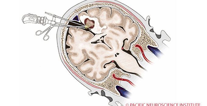
 This study evaluated an approach to reach tumors in the middle of the brain while minimizing collateral damage to the normal brain structures. By positioning the patient on his or her side, the swollen, angry brain on the side of the tumor is propped up while the normal brain gently falls away to the side due to gravity — the minimally invasive transfalcine approach. By using endoscopic visualization, we are able to successfully remove the vast majority of the tumors safely and with minimal injury to the normal brain. For metastatic brain tumors, which cause significant brain swelling, this approach is safe and successful for the majority of patients. We demonstrate the anatomy, surgical nuances and our outcomes in this article.

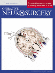
Particularly noteworthy is that the cover image for the journal was created by Pacific Neuroscience Institute medical illustrator, Josh Emerson. As a medical illustrator, Josh is a professional artist with advanced education in both the life sciences and visual communication. Collaborating with scientists, physicians, and other specialists, he transforms complex anatomical information into visual images that have the potential to communicate to broad audiences; largely including physicians, fellows, medical students, and patients. Medical illustration for the neurosciences and neurosurgery is highly complex involving the exact representation of neurosurgical anatomy and pathology. Mr. Emerson’s work has been cited in numerous neurosurgical publications and has been used as educational material in animation projects and website publications. Another example of his work is below.

Reference:
Microsurgical Endoscope-Assisted Gravity-Aided Transfalcine Approach for Contralateral Metastatic Deep Medial Cortical Tumors
Garni Barkhoudarian, MD Daniel Farahmand, MD Robert G Louis, MD Erol Oksuz, MDDanjuma Sale, MD Pablo Villanueva, MD Daniel F Kelly, MD
Operative Neurosurgery, Volume 13, Issue 6, 1 December 2017, Pages 724–731. © 2017 by the Congress of Neurological Surgeons

Garni Barkhoudarian, MD, is a neurosurgeon with a focus on skull base and minimally invasive endoscopic surgery. He has particular interest and expertise in pituitary and parasellar tumors, brain tumors, skull-base tumors, intra-ventricular brain tumors, colloid cysts, trigeminal neuralgia and other vascular compression syndromes. He is an investigator in a number of clinical trials evaluating the efficacy of various medical or chemotherapies for pituitary tumors and malignant brain tumors. For virtually all tumors and intracranial procedures, Dr. Barkhoudarian applies the keyhole concept of minimizing collateral damage to the brain and its supporting structures using advanced neuroimaging and neuro-navigation techniques along with endoscopy to improve targeting and lesion visualization.
About the Author

Garni Barkhoudarian
Dr. Garni Barkhoudarian is an expert neurosurgeon and director of the Facial Pain and Adult Hydrocephalus Centers, and Co-director of the Pituitary Disorders Center at Pacific Neuroscience Institute. His philosophy for virtually all intracranial procedures is to apply the keyhole concept of minimizing disturbance to the brain and its supporting structures.
Last updated: October 15th, 2024