Cerebrospinal Fluid Leaks
Endoscopic endonasal repair is an ideal treatment for many midline skull base CSF leaks
Cerebrospinal Fluid (CSF) Leaks occur when a defect in the coverings of the brain develops. Common causes include cranial trauma, skull base tumors and prior skull base surgery.
The most common cause of an intracranial CSF leak is transsphenoidal or transcranial surgery. Given the potential risk of meningitis and other neurological complications, urgent and effective repair of the skull base defect is essential. At the Pacific Neuroscience Institute, we have one of the world’s largest experiences in both endonasal transsphenoidal and trans-cranial repairs of skull base defects and CSF leaks. By incorporating cutting edge technology and instrumentation with proven surgical experience, we make surgery safer, less invasive and more effective.
Overview
Cerebrospinal fluid (CSF) is constantly made by the brain and reabsorbed into the blood system. On occasion, as a result of a skull fracture, weakness of the brain covering (dura), intracranial surgery or growth of a brain tumor, CSF may leak through the brain covering. This is a potentially dangerous condition that can lead to an infection of the CSF (meningitis) or of the brain itself (brain abscess).
One of the more common complications of endonasal transsphenoidal surgery is a post-operative CSF leak. As shown in Dr. Kelly’s Recent Experience, CSF leaks that occur at the time of surgery require careful repair to avoid a post-operative CSF leak. The extent and type of skull base repair depends on the size and location of the defect. Using a graded repair methodology, the chance of a post-operative CSF leak is generally 1% or less for standard endonasal removal of pituitary adenomas and other sellar tumors (Grade 1 and 2 CSF leaks and skull base defects) but is increased to 3-5% for large skull base defects (Grade 3) associated with endoscopic endonasal removal of brain tumors such as craniopharyngiomas, meningiomas and some very large invasive pituitary adenomas.
Symptoms
A CSF leak is usually associated with watery clear drainage out of the nose (after endonasal surgery or retromastoid surgery) or out of a surgical incision. If there is persistent drainage occurring within the first 1-2 weeks after surgery, you should contact your surgeon promptly. If meningitis has occurred, headache, stiff neck, fever and photophobia (sensitivity to light) may occur; other symptoms such as weakness, in-coordination and confusion may occur.
Diagnosis
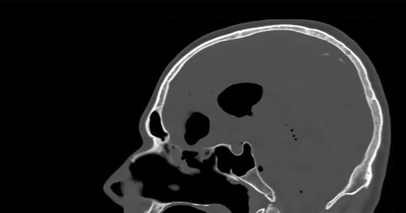
The diagnosis of a post-operative CSF leak is made by noting watery drainage out of the nose or a surgical incision. A brain MRI or CT scan may also show from where the CSF leak is originating and may reveal intracranial air (pneumocephalus). A CT scan with injection of a contrast agent through the lumbar spine (a spinal tap) can also be helpful in localizing the origin of a CSF leak and dural defect. In some instances, it may be difficult to determine if fluid from the nose or ear is actually CSF. To help determine this, a sample of the fluid can be collected and tested for a compound found only in the CSF called Beta-2 transferrin. If you have endoscopic endonasal surgery or a retromastoid craniotomy with Dr. Kelly, you will be assessed for a post-operative CSF leak prior to discharge home.
Treatment
Treatment of a CSF leak requires either direct surgical repair or in some instances use of temporary diversion of CSF through a lumbar drain or a ventriculostomy (a catheter placed in the ventricle of the brain). In some instances, both surgical repair and CSF diversion are required to effectively seal a CSF leak. For repair of large (Grade 3) anterior skull base defects associated with endonasal surgery, we utilize a pedicled naso-septal flap as part of the multi-layered repair in almost every case.
